 |
| DJM |
 | |
| Guest kitty |
Brachiocephalic vein (branches DIRECTLY off Superior Vena Cava "stem", anterior view):
 |
| Right and Left Brachiocephalic veins, respectively; DJM |
 |
| Right and Left Brachiocephalic veins, respectively; Guest kitty |
Subclavian vein (continuous with Brachiocephalic vein, i.e. "becomes" Subclavian vein; anterior view):
 |
| Left Subclavian vein, Guest kitty |
 | |
| Left Subscapular vein, Guest kitty |
Axillary vein (continuous with Subscapular vein, i.e. "becomes" Axillary vein; anterior view):
 |
| Left Axillary vein, Guest kitty |
Brachial vein (continuous with Axillary vein, i.e. "becomes" Brachial vein, anterior view):
 | |
| Left Brachial vein, Guest kitty |
External Jugular vein (anterior view):
 |
| Left Jugular vein, prior to further dissection; DJM |
 |
| Left Jugular vein, post-dissection; DJM |
 |
| Left Jugular vein; Guest kitty |
Internal Jugular vein (not visible on DJM specimen):
Pulmonary vein (Right lung):
 |
| Guest Kitty |
Inferior Vena Cava (anterior view):

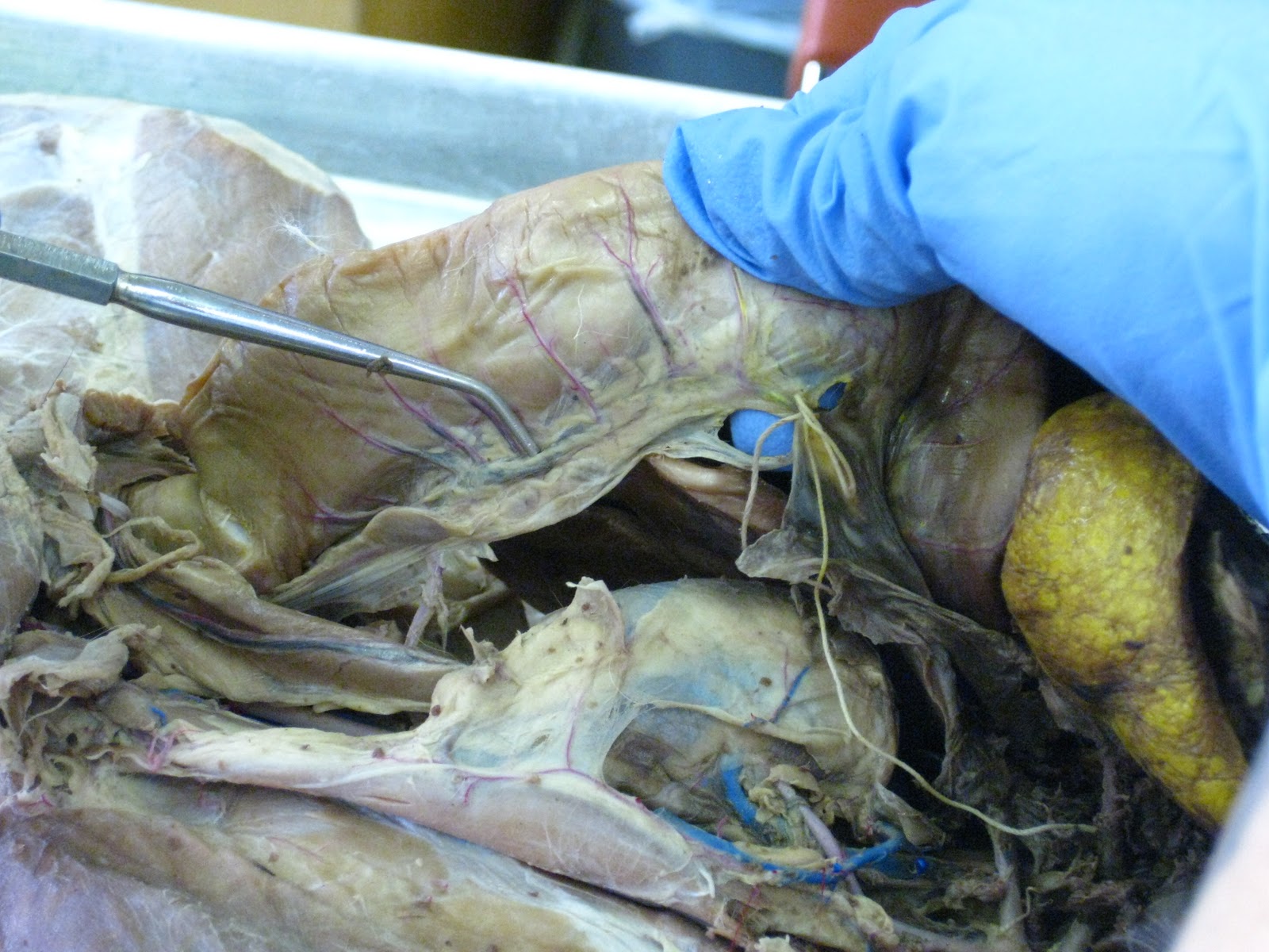
Hepatic Portal vein (does NOT branch off Inferior Vena Cava; instead, it's a vessel formed by the joining of capillaries/venules of digestive organs that transport blood, via the Hepatic Portal vein, into the liver):
 | ||
| Hepatic Portal vein (Left side of Cat); the "stem", moves blood TOWARD the liver (NOT TOWARD the heart; this is an exception to the rule of "all veins move blood toward to the heart"), formed by the UNION (anastomosis) of 2 veins: (1) gastrosplenic vein ("superior"/lateral vein), (2) superior mesenteric vein ("inferior" vein); Hepatic Portal vein stains yellow; DJM |
(1) Gastrosplenic vein (anastomoses to form Hepatic Portal vein, anterior view):
 |
| Gastrosplenic vein (upper/"superior"/lateral vein) "anastomoses" with Superior Mesenteric vein (Left side of Cat); stains yellow; Gastrosplenic vein tends to "run" with the Celiac Trunk artery; DJM |
(2) Superior Mesenteric vein (anastomoses to form Hepatic Portal vein, anterior view):
 |
| Superior Mesenteric vein (lower/"inferior" vein) "anastomoses" with Gastrosplenic vein (Left side of Cat); stains yellow; DJM |
Inferior Mesenteric vein (anterior view):
 |
| Inferior Mesenteric vein normally stains BRIGHT YELLOW and runs alongside the large intestine/colon; DJM (cat did not stain with latex very well). |
Renal Vein (anterior view):
 |
| Left Renal Vein; Renal veins are SUPER THICK and stain blue; each comes from a kidney; DJM |
(i) Common Iliac vein (1st major vein, top-to-bottom, that is continuous with the Inferior Vena Cava, anterior view):
 |
| The probe (not the forceps) is pointing to the Common Iliac vein (near the cat's groin); Common Iliac vein has 2 branches: (a) Internal Iliac vein, (b) External Iliac vein; DJM |
(a) Internal Iliac vein (branches DIRECTLY off Common Iliac vein, 2 anterior views):
 |
| The probe (not the forceps) is pointing to the Internal Iliac vein; DJM |
 |
| Both probes are pointing to the Internal Iliac vein; DJM |
(b) External Iliac vein (branches DIRECTLY off Common Iliac vein, anterior view):
 |
| The probe on the LEFT is pointing to the External Iliac vein; the probe on the RIGHT is pointing to the Internal Iliac vein for ease of reference; DJM |
(ii) Femoral vein (2nd major vein, top-to-bottom, that is continuous with the Inferior Vena Cava, anterior view):
 |
| DJM |
(iii) Popliteal vein (3rd major vein, top-to-bottom, that is continuous with the Inferior Vena Cava, anterior view):
 | |
| The probe on the RIGHT in pointing to the Popliteal VEIN; the probe on the LEFT is pointing to the Popliteal ARTERY for ease of reference. |
Great Saphenous vein (anterior view):
 |
| "Blown" Great Saphenous vein on the Professor's cat. |
Proximal Caudofemoral vein: picture not available.
Azygos vein:
 |
| The Azygous vein, which drains into the Superior Vena cava, is found on the right side of the cat. The probe is pointing to the Azygous vein in this specimen, which is (unfortunately) not well exposed; Guest cat. |
No comments:
Post a Comment